Wednesday 21 November 2012

Negotiated Study One: The Circulatory System, Further Development.


Looking at the previous design for my circulatory system and the organs that I had drawn much larger I felt that it didn't have enough detail for when I'm going to place all of the organs together. Not only that but where the organs are represented on the old design they are particularly the same shape as the organs. Then I also had the problem of when I'm going to animate really close up on the visuals there would be much to work from with the arteries, veins and capillaries because of the lack of detail. 
So I decided to redraw the heart on its own on a large scale, and then trace the outline of all the organs to then fill them in with arteries, veins and capillaries. This made the system much more complex. 
I did all of the organs separately and so will have to connect them together digitally.

A Kidney

I started to render this one but after finishing the oxygenated blood I decided that I was wasting my time as I was going to be digitally rendering it later on anyway. I think I got a bit excited to see what it would look like with colour.


The Stomach


The Pancreas


Large and Small Intestines


Left Lung


Right Lung


The Brain


The Bladde


The Heart


I'm really happy with these and am excited to get them rendered and see how I will animate it all!

Negotiated Study One: System Development Three


The Kidneys

These are two drawings that I have done for the kidneys. The second one was drawn much larger so that I can extra detail and make the little parts stronger and bolder. I'm really happy with the outcome


The Bladder

I started with a really detailed drawing of the bladder, labelling the different parts so that I can refer to both notes and drawings to know what which part is which, and what each part does!


I then simplified it. The previous drawing had more textures which  didn't include in this one as I'm going to be adding textures and colours separately a bit later on. I felt that this didn't fit in with the previous drawings that I had done as it was '3D' and so I decided to try and do it again but 2D.


This is a quick sketch when trying to make the bladder look flat.


And this is the final drawing..


The bladder is going to be kept simple like the stomach as they are both storage organs, its what happens inside them that may make them more exciting, especially the stomach!


The Brain

This is the first quick sketch of the brain, again with labels. I was pretty much really happy with this and so just drew it bigger without the labels.


The Intestines

I decided to redraw the intestines as I felt that there wasn't as much detail in them as there could be. To add more detail I again drew it much larger than previously. I'm really happy with this one and shall be my final one!


Tuesday 13 November 2012

Negotiated Study One: System Development Two


The Digestive System


I've started to explore the visuals for the digestive system, again I'm going to keep it quite true to how it looks realistically to help the audience identify what they are looking at. 

First I drew out the mouth  and throat, this is the top part of the digestive system but also the respiratory. I will probably develop this further but I can't see it becoming very detailed.


I then drew out all of the digestive organs, using my original abstract drawings for inspiration as I quite likes them. I didn't like what I did with the stomach, I was trying to show the muscle pattern but it didn't work. Another problem that I had was that I wasn't sure the shape of the small intestine looked right, in research images it was always shown with smooth walls.


So I dulled down the pattern on the stomach and small intestine but I still didn't think it looked right. The large intestine I experimented with the pattern I had used on the small intestine previously but preferred it lumpy and natural.


I the decided that it would be best to concentrate on the organs separately as I had with the systems. another thing that I had notice about the small intestine was that it wasn't as straight forward as I had been drawing it, so I made it messier. However it looked too loose and I felt that it would need to be tighter together. I was much happier with it smooth.


So I made it tighter and I was much happier although I felt that by doing so I had lost some of the messiness, especially with the bit on the right hand side.


So I tried again and this time I was happy!


I then decided to do it again but bigger so that I could get in more detail as I had with the lungs and circulatory system. The small intestines became a little looser again but I will be developing this again to correct it. 


Here I looked at the stomach and pancreas, the pancreas took my attention and so I started to develop it.


 I added detail with lots of circles to create the texture of the pancreas. As you see it here is how it would look cut in half, the veins wouldn't normally be outside. 


I went back to the stomach but decided that at the moment I wanted to keep it simple. I couldn't find any useful research images to help me add any detail and so I may come back to this. 


The same was with the liver!


Sunday 11 November 2012

Negotiated Study One: System Development


After doing the further research on the body and it's systems/organs I have decided that I will have to challenge myself and use more than four in my animation. The organs and systems that I was going to leave out previously have too much importance (everything in the body is highly important) to leave out. I've also decided that my previous designs for the systems were too simple and that it would be hard for the audience to recognise what they represent and so I'm going to use images that are more obvious as to what they are but make it abstract with the way in which I animate and how I position the shots. I won't show the entire system but rather parts of the system throughout the animation. I may at the end when the body has died 'zoom out' to show all of the systems together where the image will then fade out.

The Circulatory System


Here I drew a simple diagram of the heart, this is the clearest one that I have come across and so will probably use this version.


I then took a diagram of the circulatory system and used the diagram of the heart in it.


I then looked more closely at where the blood went round the organs. When researching it was clear that the  arteries got smaller and smaller the further they got and the veins got bigger and bigger. In the diagram above they stayed mainly the same size and so I incorporated my knowledge from my research into my next diagram.


This is much better but I feel that the proportions are all wrong, the heart is way too big compared to the other organs and so will have to redo this. I will also ad much more detail where the blood goes around the organs, I still feel that this can be improved. 


This is my improvement to this system - 


I'm a lot happier with this representation of the circulatory system, I feel that it is much more realistic. I will be making a very large version for when I animate so that I can add much more detail as I'm going to be working very close up with these images.


The Respiratory System


I drew this from a diagram of the lungs. I found that shape was wrong for the shape of the lungs and so I did it again...


Now the shape was right but I felt that it needed to be more detailed..


So I drew it bigger and added tons more branches to make it more compact and easier to recognise it as the shape of the lungs. I am really happy with this, all I need to do now is create the rest of the respiratory tract with the nose, mouth and airway down to the lungs.


Negotiated Study One: Research: The Stages of Life


The First Year

At the moment of birth the baby's lungs and circulatory system must adapt to the new, airy environment, and the other body's systems must be brought into play to sustain life outside the womb

Milk secretion begins immediately after birth. For the first few days only a yellowish fluid, colostrum, is produced by the breasts. Colostrum is of great importance to the infant because it contains maternal antibodies which help to protect the baby against infection for about three months, until it's body can produce its own defense mechanisms.

Such a the antibody - forming thymus gland in the neck which rapidly enlarges after birth.

Other important systems within the infants body rapidly mature after birth.

Enzyme systems, for example, within the intestines and the liver are soon able to digest and utilize the mothers milk. The first year is a time of rapid growth.

The infant doubles its birth weight in six months and triples it within a year.

By the age of twenty eight weeks the baby begins to eat solids.

This is the age at which the first recognizable human speech sounds begin to be produced, by contrast to the gurgles and cooing of earlier weeks.

One to Three Years

During this period the brain shows rapid growth and maturation. The most significant development is that of speech and language.

By eighteen months the child may have as many as a dozen teeth. Vocab of up to 12 words.

The two year old has about sixteen teeth and can chew efficiently. 
Vocabulary of three hundred words
Early in the childs second year the nervous system matures sufficiently to allow him to become toilet trained.


Three to Seven

The pattern of growth continues - the brain grows more slowly and the limbs and body develop quickly.

At the age of five the girls ovaries have reached their adult size, but do not function until puberty.

The 1500 word of the 4 year olds vocabulary have become 2200 on average at 5 years old.


Puberty

In both sexes the physiological changes leading to puberty occur over a period of at least two years. 

Noticeable changes usually begin in girls sometime between the ages of ten and sixteen, about two years earlier than boys.

There is a general spurt in growth hormone

At the same time there is increased secretion of hormones by the adrenals, the thyroid and the ovaries or testes. Under the influence of these hormones, sexual development accelerates.

In girls, the clearest indication of imminent reproductive ability is of course the onset of menstruation.

In the first months the loss of blood may be variable and the ovaries may not in fact be releasing fertile ova.

In males there is a growth and development of the larynx which results in deepening or breaking of the voice.


Young Adult

The aging process has begun

Loss of hearing begins in adolescence

Height begins to decrease at the age of 25.

Muscle strength begins to diminish at thirty - the percentage of fatty tissue increases.


Early Middle Years

The weight of the brain and the number of nerve cells in it begin to decline during a persons twenties.

Conduction velocity along nerves fibres decline steadily from the age of twenty 

So do the basal metabolic rate the strength of the grip, the cardiac output, liver weight and in men, frequency of intercourse. 


Later Middle Years

Physiologically, again between the ages of forty five and sixty in such a slow process that it often seems to pass almost unnoticed to the people concerned.

Consequently, in many cases the slow but relentless effects of ageing are mitigated. Sudden again or abrupt changes in health during these years is usually the result of serious illness, bereavement or some other major psychological or physical shock.


Old Age

The physiological changes which occur in old age are largely an accelerated continuation of those of the preceding years. The senses, particularly sight and hearing, all decline in activity.

Motor activity becomes impaired so that walking becomes slow.
Handwriting becomes spidery and shaky because the harmony between hand, eye and brain has lost some of it's precision.

Also seen in old age is a marked impairment of memory.
Loss of weight and reduction of height
Lowered resistance to disease and reduced efficiency of the circulatory and respiratory system.

Saturday 10 November 2012

Negotiated Study One: Research: Common Illnesses


The Common Cold

A viral infectious disease of the upper respiratory tract which effects primarily the nose.

Symptoms- cough, sore throat, runny nose fever which usually resolve in seen to ten days, some with symptoms lasting up to three weeks.
Symptoms are mostly due to the body's immune response to the infection rather than to tissue destruction by the viruses themselves.

There is no cure for the common cold.

The average adult contracts colds two or three times a year. The average child between six and twelve  times a year

A sore throat is present in about 40% of the cases and a cough in about 50%
In adults a fever is generally not present but it is common in infants and children

While a cough and fever indicates a higher likelihood of influenza in adults, a great deal of simularity exists between these two conditions

A cold normally begins with fatigue, a feeling of being chilled, sneezing and a headache, followed in a couple of days by a runny nose and cough

Symptoms may begin within 16 hours of exposure and typically peak two to four days after onset. They usually resolve in seven to ten days but some can last up to three weeks.
In children the cough last for more than ten days in 35-40% of the cases and continues for more than 25 days in 10%.

Some of the viruses that cause the common cold are seasonal, occurring more frequently during cold or wet weather.

Severe complications if they occur, are usually in the very old, the very young or those who are immunosuppressed. Secondary bacteria infections may occur resulting in sinusitis, pharyngitis, or an ear infection.
It is estimated that sinusitis occurs in 8% and an ear infection in 30% of cases.

Pneumonia can follow.


Influenza

Most common symptoms are chills, fever, sore throat, muscle pains, headache (often severe), coughing, weakness/fatigue and general discomfort.
Influenza may produce nausea and vomiting, particularly in children.

Flu can occasionally lead to pneumonia, even for persons who are usually very healthy.
In particular it is a warning sign if a child for presumably an adult seems to be getting better and then relapses with a high fever. 

Another warning sign is if the person starts to have trouble breathing

The first symptoms are chills or a chilly sensation, but fever is also common early in the infection, with body temperatures ranging from 38-39. Many people are so ill they are confined to bed for several days, with aches and pains throughout their bodies, which are worse in their backs and legs.

Symptoms- 
  • Fever and extreme cold (chills shivering, shaking (rigor))
  • Cough
  • Nasal congestion
  • Body aches, especially joints and throat
  • Fatigue
  • Headache
  • Irritated, watery eyes
  • Reddened eyes, skin (face), mouth, throat and nose
  • Petechial rash
  • In children, gastrointestinal symptoms such s diarrhea and abdominal pain (may be severe in children with influenza)
Can be hard to distinguish between the common cold  and influenza in the early stages of these infections, but a flu ca identifies by a high fever with a sudden onset  and extreme fatigue.
Diarrhea is not normally a symptom of influenza in adults.

Gastroenteritis

Is a medical condition characterized by inflammation of the gastrointestinal tact that involves both the stomach and small intestine, resulting in some combination of diarrhea, vomiting and abdominal pain and cramping.
Transmission may occur due to consumption of improperly prepared foods of contaminated water via close contact with individuals who are infectious.

Gastroenteritis typically involves both diarrhea and vomiting, or less commonly, presents with only one or the other.
Signs and symptoms usually begin 12-72 hours after contracting the infectious agent

If due to a viral agent, the condition usually resolves within one week.
Some viral causes may also be associated with fever, fatigue, headache and muscle pain.

Children infected with this usually make a full recovery within three to eight days.

There are many causes of gastroenteritis; most numerous cases are caused by viruses, followed by bacteria and other agents.

Food Poisoning

Food poisoning is an illness caused by eating contaminated food - most people will get better without the need for treatment.

In most cases, the food that causes the illness has been contaminated by bacteria, such as salmonella or E.coli , or a virus, such as the norovirus.

The symptoms of food poisoning usually begins one to three days after eating contaminated food. They include feeling sick, vomiting, diarrhea and stomach cramps.

Foods that are particularly vulnerable to contamination if they are not handled, stored or cooked properly include raw meat, and poultry, 'read to eat' foods such as cooked sliced meats, pate, soft cheeses and prepacked sandwiches, dairy products, such as eggs and milk.

Symptoms of the food poisoning depend on the type of contaminant and the amount eaten. The symptoms can develop rapidly within 30 minutes, or slowly, worsening over days to weeks.

Usually food poisoning is not serious, and the illness runs its course in 24-48 hours


Tonsilitus

Is an acute swelling and irritation (inflammation) of the tonsils. Tonsilitus is caused by a bacterial infection or a viral infection.

Tonsils are two glands located in the back of the throat. They belong to the lymphatic system and the immune system and help to protect the body from upper respiratory infections.
Especially important n young children
Can occur to anyone with tonsils but is most common in children.

Th throat and tonsils become red, swollen and white patches of pus may appear on the tonsils.
Other symptoms include pain when swallowing, difficulty swallowing, headache fever and swollen glands.

Contagious and spread when an infected person talks, coughs or sneezes. This shoots droplets contaminated with bacteria or a virus into the air where hey can be breathed in by others.


Sinusitis

Inflammation of the paranasal sinuses, which may be due to infection, allergy, or autoimmune issues. Most cases are due to a viral infection and resolve over the course of 10 days.
It is a common condition; for example, in the US more than 24 million cases occur annually.

It is defined as an inflammation of the mucous membrane that lines the paranasal sinuses and is classified chronologically into several categories.

Acute sinusitis is very common. Roughly 90% of adults have had sinusitis at some point in their life.

Symptoms- 
  • Headache/facial pain or pressure of a dull, constant, or aching sort over the affected sinuses in common. This pain is typically localized to the involved sinus and may worsen when the affected person bends over or when lying down. Pain often starts on one side of the head and progresses to both sides.
  • This may be accompanied by thick nasal discharge that is usually green in colour.
  • Often a localized headache or toothache is present, and it is these symptoms that distinguish a sinus-related headache from oth types of headaches, such as tension or migrane headaches
Sinus infections can also cause inner ear problems due to the congestion of nasal passages. This can be demonstrated by dizziness, 'a pressurized or heavy head', or vibrating sensations in the head.


Ear Infections

Otitis - a general term for inflammation or infection of the ear.
Otitis externa involves the outer ear and ear canal. In external otitis the ear hurts when touched or pulled.
In otitis media or middle ear the ear is infected or clogged with flud behind the ear drum, in the normally air filed middle ear space. this very common childhood infection sometimes require a surgical procedure called myringotomy and tube insertion.
Otitis interna involves the inner ear. The inner ear includes sensory organs for balance and hearing. When the inner ear is inflamed, vertigo is a common symptom.

Ear infection is common in children, but can occur at any age. The main symptoms are ear ache and feeling unwell. Painkillers are the main treatment.

The small space behind the eardrum in the middle ear is normally filled with air. It is connected to the back of the throat by a tiny channel called the Eustachian tube.

The middle ear space sometimes becomes filled with mucus(fluid) often during a cold. The mucus may then become infected by bacteria or viruses.
Children with glue ear who have mucus behind their eardrum are more prone to ear infections. Sometimes an ear infection occurs 'out of the blue' for no apparent reason.

Symptoms- 
  • Earache is common, but does not always occur
  • Dulled hearing may occur for a few days
  • Fever is common
  • Children may feel sick or vomit
  • Sometimes the eardrum perforates (bursts). this lets out infected mucus and the ear becomes runny for a few days. As the pain of earache is due to a tense eardrum, a burst eardrum often relieves pain. A perforated eardrum usually heals within a few weeks after the infection clears.
Most bouts of ear infection will clear on their own without treatment within 2-3 days.


Pharyngitis

Inflammation of the throat. In most cases it is quite painful, and it is the most common cause of a sore throat. 
Pharyngitis can result in very large tonsils which cause trouble swallowing and breathing. Pharyngitis can be accompanied by a cough or fever.

Pharyngitis is a ype of inflammation, most commonly caused by an upper respiratory tract infection.
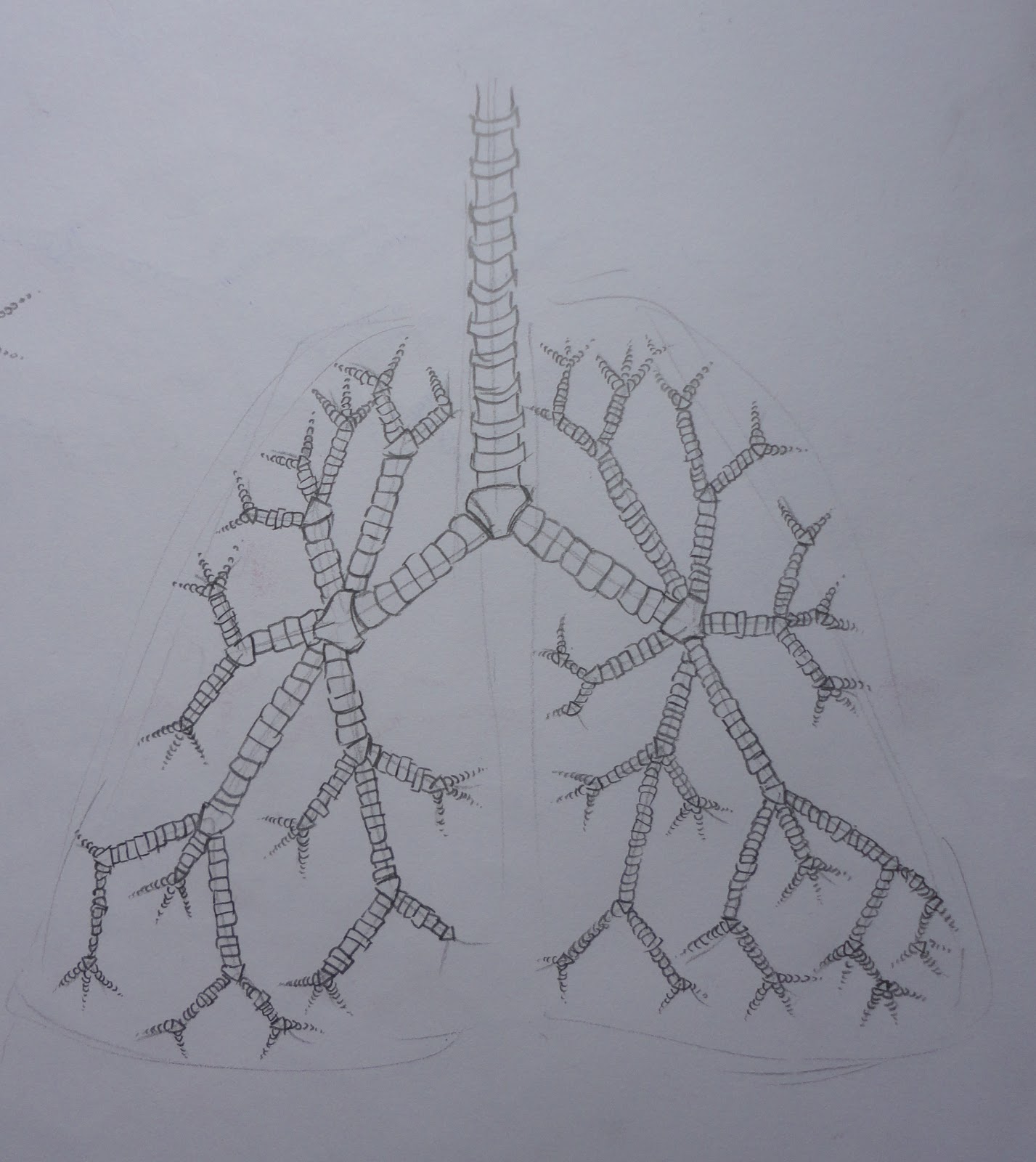
Pneumonia

Pneumonia is an inflammatory condition of the lung - specially affecting the microscopic air sacs (alveli) associated with fever, chest symptoms and a lack of airspace o a chest xray. Pneumonia is typically caused by an infection but the are a number of other causes.

Typical symptoms include cough, chest pain, fever and difficulty breathing.

People with infectious pneumonia often have a production cough, fever accompanied by shaking chills, shortness of breath, sharp or stabbing chest pain during deep breaths, confusion, and an increased respiratory rate.
In the elderly confusion may be the most prominent sign. The typical signs and symptoms in children under five are fever, cough, and fast or difficult breathing.

Due to primarily infections, with less common causes including irritants and the unknown.


Shingles

Herpes Zoster, commonly known s shingles and also known as Zona, is a viral disease characterized by a painful skin rash with blisters in a limited area on one side of the body, often in a stripe.
The initial infection causes the acute illness chicken pox which generally occurs in children and young people. Once an episode has resolved, the virus is not eliminated from the body but can go on to cause shingles - an illness with very different symptoms - often many years after the initial infection.

Years or decades after a chicken px infection, the virus may break out of nerve cell bodies and travel down nerve axons to cause viral infection of the skin in the region of the nerve. the virus may spread from one or more ganglia along nerves of an affected segment and infect the corresponding dermatone (an area of skin supplied by one spinal nerve) causing a painful rash. Although the rash usually heals within two to four weeks, some sufferers experience residual nerve pain for months or years, a condition called postherpetic neuralgia.

The earliest symptoms include headache, fever and malaise are non specific, and may result in an incorrect diagnosis. these symptoms are commonly followed by sensations of burning pain, itching, oversensitivity or pins and needles, tingling, prickling or numbness. The pain maybe mild.

Friday 9 November 2012

Negotiated Study One: Further System Research: The Kidneys and The Brain and Central nervous System


Kidneys: The Filtration Units


  • The urinary system is concerned with the formation and elimination of urine
  • In an adult, more than 2500 pints of blood pass through the kidneys each day
  • Blood enters via the renal arteries and is filtered to remove most of the waste products of metabolism.
  • Seven pints of filtration are produced every hour. Purified blood returns to the body circulation via the renal veins
  • The filtering process is carried out by more than two million tiny kidney units, or nephrons, which produce a highly concentrated solution of chemicals known as urine, which is harmful to the body if allowed to remain.
  • Urine flows from the nephrons, first into the funnel shaped renal pelvis and then into the ureter. 
  • Waves of muscular contraction passing down the ureters push the urine into the bladder.
  • With continuous filling, the bladder, a muscular bag, expands until it holds about one pint of fluid. 
  • A circular band of muscle around the neck of the bladder, the sphincter, controls the release of urine from the body.


  • Themost important route of excretion is through the kidneys - the paired filtration units lying at the back of the abdomen. 
  • These produce a constantly adjusted trickle of urine which is voided when the bladder becomes uncomfortably full.
  • Urine, a solution in water of urea, salts and other soluble wastes, is produced by the kidneys from the blood that enters them, under high pressure, through the renal arteries.
  • The renal arteries are large side branches of the aorta and together receive about a quarter of the volume of blood pumped out by the heart at each beat.
  • All of the blood in our bodies passes through the kidneys about twenty times every hour, but only one fifth of the plasma is filtered into the kidneys tubules.
  • Each kidney is roughly the size of a child's fist
  • Seen cut open it has an outer pale layer called the cortex, and a darker core, densely packed with blood vessels that divide and subdivide, called the medulla
  • On the medial aspect of each kidney lies a urine collecting cavity, the renal pelvis, leading down through a thin walled tube, the ureter, into the distensible bladder
  • Each kidney has about one million separate filter units, called nephrons. A nephron consists of a cup shaped structure, Bowmans capsule, which encloses a know of capillaries, known as glomerules that carries blood from the renal artery, and a renal tubule.
  • Each tubule is between one and two inches long, and loops, rather like a trombone, in and out from the cortex, where the capsule lies into the medulla
  • The tubules unite into collecting ducts, and the ducts empty their contents into the renal pelvis.

  • As blood under pressure passes through each glomerus, water, dissolved salts, sugar, urea and many other small molecules are forced through the walls of the capillaries and into the capsule.
  • The blood, now more concentrated because it has lost about one fifth of its plasma, passes from the glomerus into a network of fine vessels surrounding the loops of each tubule, and will eventually rjoin the general circulation through the renal vein.
  • Before it does however, another important process must be carried out.
  • Although the kidney is described as excreting water, it has in fact a duty to conserve water.
  • our bodies need water as a medium in which every biochemical process can take place . Only if we were water logged could water be said to be a waster material with a nuisance value.
  • It is simple that a solvent is needed for true wastes, to remove them from the body, that large amounts of water pass into the kidney tubules
  • Nearly all the water i, therefore, recovered from the filtrate in its journey through the loops of the renal tubule, passing back into the bloodstream through the tubule walls into the networks of blood vessels.

  • At the same time by process of chemical transfer through the tubule walls, sugar and other materials that the body needs, but which have passed into the filtrate because of the small size of their molecules, are also conserved by reabsorption into the blood.
  • So efficient is the reabsorption of sugar, for example, that not a trace is normally detectable in the urine. 
  • After passing down the ureters, the urine is stored in the bladder, to be discharged through the urethra to the outside when convenient.


The Brain and Central Nervous System


  • Monitoring and controlling every aspect of the body's activities, no matter whether we are awake or asleep, is the vast and complex communications network called the nervous system.
  • At the very heart of the network is the central nervous system, consisting of the brain and spinal cord. 
  • And weaving through and penetrating to every near and outlying region of the body are the almost countless branches of the peripheral nervous system.
  • The all important brain, a densely packed mass of nervous tissue weighing nearly three pounds, and the spinal cord, continuous with the brain.
  • Acting as channels of communication between the central nervous system and the rest of the body are the afferent or sensory receptors, and the efferent, or motor, nerves, which carry outgoing messages to effector structures such as the voluntary muscles of the limbs.
  • Sensory and motor nerves of the peripheral system leaves the spinal cord separately, between the vertebrae, but then unite to form thirty one pairs of spinal nerves, in which there are both sensory and motor nerve firbres
  • Branches of the spinal nerves spread out to reach all parts of the body surface and all skeletal muscles


  • Leaving the brain directly are twelve pairs of cranial nerves, each passing through the separate aperture in the skull
  • There are more than ten thousand million nerve units, or neurons, in the brain, but even these account for only one tenth of the brain cells. 
  • The remainder are surrounding and supporting cells called the neuroglia or just glia.
  • Together the neurons and glia make soft, jellylike tissue; without the support of the surrounding skull it would distort and sag.

Negotiated Study One: Further System Research: Metabolism, The Circulation of Blood and Respiration


Metabolism


  • During his lifetime the average man eats about fifty tons of food and yet maintains a weight of about 160 pounds
  • Even the most obese person retains in his body only a minute fraction of what he eats.
  • Most of the food we absorb into out bodies through the process of digestion must sooner or later pass out again
  • If we eat more food than required to provide energy or for tissue growth and repair, the excess is stored in adipose tissue as fat. This fat acts as our energy store and as an outer insulating layer
  • The advantage of storing excess food as fat is that it is a compact, energy rich compound and all that is needed for its synthesis is acetyl-coA and appropriate enzymes


The Circulation of Blood


  • We have a complex transport system that pumps and circulates blood around the body
  • Blood has  special properties that enable it to take up oxygen in chemical combination in regions where it is plentiful -  those in contact with the outside air - and then, during circulation, to release it where oxygen is being used up and is not so plentiful - deep in the bodies tissues
  • During circulation, blood can also transport carbon dioxide in the opposite direction, from the tissues where it is produced to the outside air, so that the result is an exchange of the two gases. 
  • We have developed a special internal surface - the linings of the lugs - through which such an exchange can take place readily.
  • the total surface area provided by the lungs is about forty times that of the skin.
  • Circulating here, beneath the moist, thinner - than - paper lining blood can come into very close contact with the air that we breathe, and carry out the exchange. 


  • Blood circulating in the walls of the intestines picks up water and dissolve food substances that are destined for distribution and use as fuel and construction materials throughout the whole body.
  • Some but very little of the waste matter produced by living cells does in fact find its way out of the body through the skin, such as sweat. The components of sweat, including water and salts, are derived from the bloodstream. By far the greater proportion of unwanted material leaves the body in the urine, a solution filtered by the kidneys from the blood circulating through them
  • This is the main route of excretion

  • The essential function of the blood, then, is to transport substances as it circulates.
  • The human body has a pump, the heart, and a vast network of blood carrying vessels that penetrates to every corner of the body, servicing and bathing with fluid every cell in every tissue
  • Arteries and veins are the largest tubes of the circulatory system, and the major routes by which the blood travels to all parts of the body from the heart, and returns to it for recirculation.
  • Arteries all carry blood away from the heart,  and veins all carry blood toward the heart.
  • The blood in arteries is under high pressure, and the vessel walls are appropriately thick, muscular and slightly elastic.
  • The surge of pressure that follows each contraction of the heart causes the arterial walls to yield slightly, smoothing out the blood flow. 
  • This momentary expansion echoing every heart beat, is felt as a pulse where any large artery runs near the surface of the body
  • The largest artery in the body is the aorta, nearly one inch wide, that arches, like the handle of a walking stick, up and backward from the left ventricle of the heart to pass down infront of the spine, giving off branches in its course to supply all parts of the body. 
  • Arteries branch into smaller vessels, and at their ends become arterioles, which form a network in the skin, in the muscles and in the walls of the arterioles enable them to contract and so regulate the blood flow in particular regions according to needs. 
  • Arterioles in turn divide into minute capillaries, tubules averaging only on twenty fifth of an inch in length, and a hundredth of that in diameter.
  • Capillaries have a wall only one cell thick, through this wall materials pass into and out of the blood.
  • On the return side of the circulation, capillaries join up with the smallest branches of the veins, the venules, and the venules unite to form wider and wider collecting vessels until they becomethe major veins that return blood to the heart.
  • Veins have thinner walls than arteries and the pressure in them are lower. 
  • They expand and collapse to adjust to the blood volume within them. 
  • The massaging effect of nearby muscles, and the presence of simple cup shaped valves in the larger veins, helps to prevent blood from collecting in the lower parts of the body. 


  • The basic circulation, then, is from heart to artery, to arteriole, to capillary, to venule, to vein and back again to the heart.
  • But in fact, because blood must pass through the lungs to pick up oxygen and release the carbon dioxide it has gathered from tissues, there is a double circuit. 
  • Instead of being pumped directly around the body again, blood returning from the tissues is sent through capillaries in the lining if the lungs and taken back to the heart through the pulmonary veins
  • Now the circuit can start again

The Heart: A Muscular Pump

  • Because there is a double circulation, blood in effect travels in a figure eight circuit.
  • The heart lies at the point of cross over and can be regarded as two linked pumps.
  • Because the systemic circulation is so large, the left ventricle has the greatest muscle mass; the apex beat is due to its forceful movement
  • Both atria and both ventricles contract at the same time in rapid succession.
  • This period of ventricular contraction is called systole, and is followed by a relaxation or recovery period called diastole.
  • Systole and diastole are both very short.
  • he heart goes through about seventy cycles of systole and diastole every minute, but its rate can be more than doubled to keep up with the body's oxygen needs during violent exertion.
  • It is the valves of the heart that turn it into a pump. They are one way valves, which means that they shut automatically of blood attempts to move against the proper direction of flow.
  • The caliber of the arterioles is constantly changing under the influence of the autonomic nervous system, according to the body's needs, and the rate and force of the heartbeat are also adjusted by hormones, particularly adrenaline, which circulates in the bloodstream.


Respiration


  • Every minute we breathe in and out between ten and fifteen times
  • Brain cells are particularly sensitive to oxygen lack; starved of the gas for just a few minutes, they dies and cannot be replaced. We must breath to stay alive. 
  • Two systems in particular are responsible for supplying all the body's cells with oxygen and, at the same time, for disposing of the waste carbon dioxide and water resulting from the cells metabolism.
  • These are the respiratory system, of airways and lungs; and the circulatory system, of heart, arteries, capillaries and veins.

  • Air enters and leaves the body through the nose and mouth.
  • At the entrance to the nasal cavities, paired passageways separated by a bone sectum, are the nostrils.
  • Coarse hairs act as a trap of large dust particles that may be inhaled
  • Within the nasal cavities the air that is breather in passes over the thin, glistening membrane lining the walls, and is warmed and moistened. 
  • The mucus membrane is richly supplied with blood vessels and is capable of raising the temperature of the air from near freezing, on a cold day, to almost that of the body by the time it reaches the back of the throat.
  • Warm, moistened and filtered, the air now passes into the pharynx, the cavity behind the nose and mouth.
  • The air is sucked down into the larynx, the box like structure that supports the vocal chords and then goes into the lower respiratory tract.

  • During swallowing, the entrance to the trachea is momentarily closed by a flap called the epiglottis to prevent food entering but the tube itself is always kept open by c-shaped struts of cartilage.
  • At its lower end, the trachea divides into the bronchi, one of which enters each lung, there to divide and subdivide and lead eventually to several hundred million air sacs, called alveoli.
  • Here the exchange of gases between the inhaled air and the blood flowing in networks of capillaries takes place.

  • The lungs fill most of the chest cavity, they are sponge like structures that are inflated and deflated by the movements of the chest wall and the rise and fall of the diaphragm- the sheet muscle that divides the chest from the abdomen below. 
  • Each lung is enclosed in a double layer of fine membrane called pleura; the outer pleura is closely attached to the walls of the chest cavity and to the upper surface of the diaphragm, and the inner pleura is attached to the lungs.
  • During normal breathing it is mainly the diaphragm that does the work. 
  • As the muscle contracts, stimulated by the slow rhythmic impulses carried by nerves from the brain, the volume of chest cavity is increased. 
  • At the same time the ribs, hinged where they join the spine, and link to the sternum, or breastbone, at the front, move upward and outward, pulled by the muscles between them, the intercostal muscles.
  • As the volume of the chest increase, so the pressure within drops, and air is suck into the lungs through the trachea, restoring the pressure. This is inspiration

  • Expiration is normally an effortless process. The diaphragm and intercostal muscles relax, and the elasticity of the spongy lung tissue forces air out as the volume of the chest cavity decreases.
  • With muscular effort, considerably more air can be squeezed from the lungs. 
  • The respiratory system is able to adapt to changes in the body's needs. As the rate at which energy is used up increases, so the rate and depth of respiration are automatically adjusted.
  • The quantity involved is called the tidal volume
  • During physical exertion as when you are running for a bus, the amount of air needed may be some twenty times as much as during rest.
  • The rate of respiration may be more than doubled , and, at the same time, the amount of air per breath is greatly increased.
  • With effort up to five pints of more air can be taken in during inspiration
  • Breathing is normally an involuntary activity, but it can, at least within certain limits, be controlled voluntarily.

  • Coughing, sneezing and hiccuping, sighing, yawning, crying and laughing are all unusual forms of respiration and occur in response to physical or emotional triggers.
  • Strong smells or dust particles, for example, can irritate the nasal lining and cause sneezing, while the cough is a response to the presence of irritant material, such as dust in the bronchi, or to excess mucus produced in response to infection or noxious fumes.
  • Hiccups result from sudden, sharp contractions of the diaphragm, stimulated by the vagus, a nerve which is also involved with the digestion. That is why hiccuping can be associated with eating certain foods.
  • Crying and laughing are both long inhalations followed by a burst of short, staccato exhalations and are respiratory reactions to emotional stimuli.


Negotiated Study One: Further System Research - The Digestive System and The Liver


The Digestive Tract


"Man is a machine fueled by food. If food is to be utilized it must be broken down into molecules small enough to be absorbed. 
This is the duty of the digestive tract, a 26ft long tube from mouth to anus in which food is subjected to a coordinated thermal, mechanical and above all, chemical assault called digestion. 

Mouth

  • Food enters the mouth where it is warmed or cooled to the optimal temperature for digestion. 
  • Solid food is torn and ground by the teeth into small pieces, which are easier to swallow and digest.
  • Dry food is now mixed and lubricated with saliva to ease its passage through the esophagus
  • Each day the salivary glands produce about three pints of saliva which contains the starch-digesting enzyme ptyatin. This however has little chance to act before food is swallowed.


Esophagus

  • Food leaves the mouth when we decide to swallow, a process which once begun is beyond conscious control.
  • When the ball of food leaves the mouth it passes through the esophagus, where powerful waves of muscular contraction squeeze it down to the stomach.


The Stomach

  • The adult stomach is a distensible sac which can hold up to two and a half pints
  • It serves to store food so that it can be slowly pushed on to the duodenum at the rate required, while the gastric juice it produces starts the break down of the proteins in the food and kills any contaminating bacteria. 
  • The activities of the stomach are controlled by hormones and nerves
  • The sight, smell and even the thought of food will stimulate the production of the gastric juices
  • There are three major components of the gastric juices - digestive enzymes, hydrochloric acid and mucus
  • Digestive enzymes are compounds which cause the chemical breakdown of food.

  • The rate at which food leaves the stomach is controlled by its chemical composition
  • Carbohydrate rich food leaves the stomach in a few hours
  • Protein rich food is retained longer
  • While it takes many hours before fatty food is gone
  • Proteins and fats, which are more difficult to digest than carbohydrate s, cause the duodenum to release into the blood varying amounts of the hormone entrogastrone, which reduces gastric activity, so that the duodenum receives food at the rate it can deal with, rather than at the rate the food is eaten.


The passage of food through the stomach


Duodenum

  • As soon as acid is detected, the hormone secretin is released. This stimulates the pancreas to secrete a watery alkaline juice that neutralizes the corrosive hydrochloric acid.
  • The hormone pancreozymin the causes the pancreas to start the somewhat slower production of enzymes to break down proteins and start the digestion of the carbohydrates, the fats and the nucleic acids.
  • The presence of fats causes the duodenum to release yet another hormone, chlocystokinin, which makes the  gall bladder contract. As a result, bile, a thick green alkaline solution of salts and pigments made in the liver and stored in the gall bladder, is poured out and mixes with food. 
  • Bile salts are necessary for the efficient digestion of fats, which they emulsify so that they are more easily acted on by enzymes.

  • By the time it leaves the duodenum almost anything we have eaten looks the same - a watery mixture of partly digested food, enzymes and other excretions.
  • This mixture still needs further processing before it can be absorbed
  • Both of these processes occur in the next and longest part of the small intestine

  • The logic of absorption and digestion is best understood when it is realized that a human being is a kind of fancy shaped doughnut, whose digestive tract is the holes in the middle

The Small Intestine
  • When the chyme reaches the small intestine, although some food substances are ready to be absorbed, further processing is required for most. 
  • Food protein has already fragmented into chunks called peptones and these are now broken into the absorbable amino acid
  • Sugars and starches, similarly fragmented in the duodenum, are broken down into individual monosaccharides from which are built.
  • Fats emulsified by the action of bile are split into particles which can be absorbed. 
  • All this is achieved by the enzymes contained in the intestinal secretion
  • About five pints of this secretion in produced each day, but only in response to food. 
  • The food is slowly pushed along the small intestine by rhythmic contractions, which also break up any remaining lumps.
  • As this final breakdown of food is occurring the end produced are being absorbed across the intestinal wall into the body.
  • the small intestine is a narrow tube which is about twenty one foot long and the amount of absorption required is enormous
  • the volume total of food, drink and gastrointestinal secretions is some two and a half gallons per day pf which only about one fifth of a pint is finally lost in the feces. 
  • To facilitate the absorption of nutrients as well as water, the wall of the small intestine is folded to increase its surface area
  • Not only do broad, spirally arranged folds create a series of permanent ridges down its length, but also these are in turn covered on the inner wall by minute finger like projections called villi.
  • The villi are just visible to the naked eye and are said to give the intestine a velvety appearance
  • The villi increase the absorptive surface of the intestine by about five times what it would be if smooth, just as a piece of velvet has a mush larger surface area than a piece of smooth cotton cut to the same dimensions. 

  • Villi themselves are further subdivided their surface being covered in the microvilli, so small that only the most powerful microscope can detect it

  • One square inch of intestinal wall is covered by something like twenty thousand villi and more than ten thousand million microvilli

  • Although this enormous surface makes the absorption of food highly efficient, it also increases the area of cells exposed to wear and tear

  • Once absorption is complete, a watery mix containing mainly fibrous waster, indigestible cellulose, some salts, and unwanted breakdown products remain, and this mow passes through the ileocecal valve into the large intestine.

The Large Intestine

  • On a normal western diet, about twelve ounces of chyme enters the cecum every day. The continuing reabsorption of water reduces its weight by two thirds, forming the feces.
  • These weight however, are extremely variable - much reduced on a refined diet and considerably increased by the consumption of roughage
  • Most of the large intestine is colonized, permanently and harmlessly, by bacteria which use the indigestible residues as nutrients and my synthesize some of the bodys victim supplies
  • there is a continual loss of these bacteria into the feces
  • Like the stomach, the large intestine, particularly the descending colon and the rectum, has use as a store
  • Although the presence of increasing amounts of feces causes a reflex desire to defecate, the large intestine can store considerable amounts of fecel matter

The Liver

  • The  liver is a central chemical processing plant of staggering complexity. Weighing three pounds, it is the largest single gland in the body and with more than five hundred functions so far indentified, it is easily the most versatile.
  • Unlike other organs, the liver is served by two distinct blood supplies. The heptatic artery supplies t with fresh oxygenated blood, while the hepatic portal vein transports blood to it for processing.
  • This portal vein supply comes from the stomach and intestines carrying absorbed nutrients which the liver extracts, processes and stores, and from other main abdominal organs - the spleen, the gall bladder and the pancreas.
  • When the portal vein enters the liver it branches repeatedly, its smaller branches, running with branched of the hepatic artery and bile ducts along the connective tissue - lined tubes that surround the liver lobules
  • The lobules are tiny filters of which is, in effect a chamber of hepatic cells honeycombed by an interconnecting maze of sinuses, or caverns, that lead to a central draining vein.
  • The central veins, in turn, join, finally leading to the hepatic vein that takes blood from the liver. 
  • The cells of the lobules are washed by arterial blood, bringing them necessary oxygen, and portal blood, from which they remove nutrients, bacteria and old red blood cells.
  • At the same time, into this cascade of blood passing through the microscopic sinuses, the secrete glucose, proteins, vitamins, fats and most of the other compounds required by the body. 

  • Running between the individual cells that line the sinuses is yet another network of tunnels, much smaller than the sinuses, which drains outward, carrying bile toward the local branch of the bile duct, at the edge of the lobule.
  • Bile, a solution of organic compounds in water, is produced by the hepatic cells. Some of the contents of the bile pigments that give it its green colour, cholesterol and bile salts produced from cholesterol are, in part, waster products from the breakdown of old red blood cells.
  • The economy of the body is such that the waste products are sued as essential substances for the digestion and absorption of fats in the intestine.
  • Part of the bile salts and cholesterol in the bowel is absorbed and recycled and part is lost in the 'drain' of the intestine, with the pigments in the bile contributing to the colour of the feces.
  • Eventually, the pint of bile produced each day reaches the gall bladder, a small muscular sac underneath the liver, where it in stored until it is needed in digestion

  • When hemoglobin is broken down in the liver much of the iron that it contains it reutilized
  • Excess amino acids from hemoglobin, or, more generally, from all the other proteins that are broken down, are either used by the versatile hepatic cell for building new proteins or further broken down to provide energy.
  • Poisonous ammonia produced during this process is converted to urea, a non toxic compound, and returned to the blood for transport to the kidneys.
  • From there it is excreted as urine.

  • Perhaps what is most surprising of all is that the  site of this immense chemical plant - factory, warehouse and sewage works, is the simple hepatic cell
  • The liver has another property that makes it different from most of the rest of the body. If it is damaged, or part is removed, it can regrow to a large extent to continue its work as before.


  • The liver is the largest organ in the body
  • Liver tissue is composed of a compact mass of multisided units, the hepatic lobules.
  • Each lobule consists of a central vein surrounded by plates of liver cells.
  • The liver receives blood from two sources; 80 percent which carries digested food materials, arrives from the intestines via the portal vein; the remaining 20 percent is oxygenated blood from the heart, which enters through the hepatic artery.
  • An exchange of materials takes place between liver cells and the blood, which then passes into the central veins and returns to the general body circulation via the hepatic vein.
  • When an adult is at rest, about two and a half pints of blood flow through the liver each minute.